IUFRO Working Party 7.02.02 FOLIAGE, SHOOT & STEM DISEASES
June 13-19, 2004, Oregon, USA
P.16
Magnetic Resonance imaging of xylem dysfunction in Quercus crispula infected with a wilt pathogen, Raffaelea quercivora
by K. KURODA1, Y. ICHIHARA2, Y. KANBARA3, T. INOUE4, and A. OGAWA4
1: Kansai Research Center, Forestry and Forest Products Research Institute
Momoyama, Fushimi, Kyoto 612-0855, Japan
2: Tohoku Research Center, Forestry and Forest Products Research Institute
3: High Field Magnetic Resonance Imaging Research Institute, Iwate Medical University
4: Department of Neurosurgery, Iwate Medical University School of Medicine
Email: keiko@affrc.go.jp
http://cse.ffpri.affrc.go.jp/keiko/hp/kuroda-e.html
Summary
The large mortality rate of deciduous oaks, Quercus serrata and Q.
crispula, has been a serious problem for the past two decades in
Japan. The pathogen of this wilt disease, Raffaelea quercivora,
enters the trunks by mass attacks by an ambrosia beetle, Platypus
quercivorus. Physiological and cytological investigations of
infected trees revealed that widespread discoloration and serious
dysfunction had occurred in sapwood prior to the wilt. The
present report discusses the mechanism of wilting based on the
non-destructive observation of xylem dysfunction using the MR imaging
technique after the inoculation of three-year-old Q. crispula trees
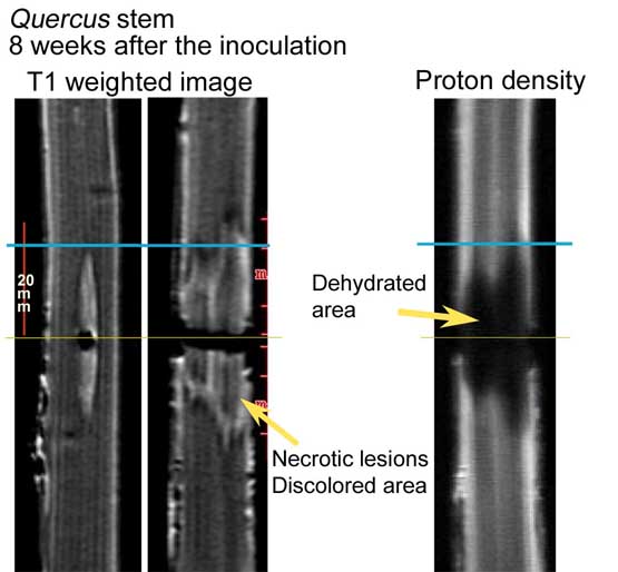
with the pathogen. The MR imaging shows water-containing tissues
as high intensity as a result of the behavior of proton.
Conductive vessels in healthy oak trees were recognized as whitish
areas on the MR images. By one week after the inoculation with R.
quercivora, the area ca. 1cm above and below the inoculated sites
showed low intensity and was looked darker. Eight weeks after the
inoculation, the area of low intensity had reached about 2cm above and
below the infection site. Anatomical observations after the MR
imaging revealed that water conduction had stopped and there was
desiccation in the darker areas. T1 weighted images, which is
used to detect the presence of fat or protein for medical purposes,
showed the necrotic and discolored xylem area as high intensity.
Some substances produced by the activity of the pathogenic fungus might
be detected on T1 weighted images.