IAWA Journal, Vol. 23(4), 469-470, 2002
Abstracts 5th Pacific Regional Wood Anatomy Conference
(Yogyakarta)
Analysis of NMR-CT images to detect the xylem dysfunction and
lesions in tree trunks
Keiko Kuroda1, Y. Kanbara2, T. Inoue2, and A. Ogawa2:
1 Forestry and Forest Products Research Institute,
Kansai Research Center, Kyoto 612-0855, Japan;
2 High Field Magnetic Resonance Imaging Research
Institute, Iwate Medical University, Takizawa-Mura, Iwate 020-0173,
Japan.
The capability of xylem water conduction, which is
an important factor related to the tree health, is difficult to assess
on the living trees. NMR-CT (MRI: Magnetic Resonance Imaging)
developed for a medical checkup was tested for the purpose to detect the
water distribution and pathological incidences in tree trunks.
Detection of tree's internal phenomena without cutting specimens into
pieces will develop the researches in the field of tree physiology,
functional anatomy, and pathology. As materials, Pinus and Quercus
species are selected. In addition to healthy trees, trees
inoculated with pathogens of pine wilt (Bursaphelenchus xylophilus)Å@or
of the wilt disease of oak trees (Raffaelea quercivori) were
selected. Trunks (3to 4cm in diameter) cut about 40cm in length or
a whole tree (1cm in diameter) was scanned.
NMR images from a healthy Pinus tree demonstrated
the areas filled with water showed white: cambium and most part of early
wood. Latewood and compression wood looked darker in September,
probably because the narrow tracheids were dehydrated (embolized) for
the drought during summer and dysfunctional. Pith was black.
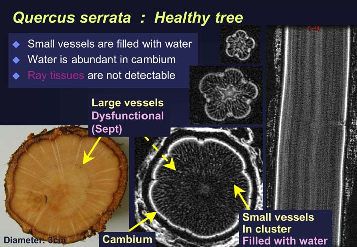
NMR cross-sections of healthy Quercus trees indicated cambium and
clustered small vessels were white and filled with water. Large
vessels were not filled with water and looked darker in September.
In the trees infected with pine wilt, dysfunctioned tracheids emerge
within a short period after the invasion of the pathogen. So far
the dehydrated areas had been checked by tissue dissection. By the
NMR imaging of a whole tree, the dysfunctioned area was shown as
pitch-black. In Quercus trees infected with R. quercivori but not
wilting, dysfunctioned xylem area also appeared darker. In
addition, the lesions where the fungal hyphae distribute and parenchyma
cells are necrotic showed whitish. Some substances increased by
the fungal activities may be detected. Three-dimensional images of
trunks can be reconstructed from cross-sectional MRI data taken at
intervals of several millimeters through 120cm. These images are
helpful to grasp the extent of dysfunctional xylem and lesions.
NMR-CT is a powerful tool to analyze the distribution of water in the
trees and may be useful to detect the areas affected by the pathogenic
microorganisms.
Back
Back (Japanese)