KURODA, Keiko
Jpanese version is here
.
December 17, 2008
Forestry
and Forest Products Research
Institute
kansai Research Center
Forest health Research Group
Momoyama, Fushimi, Kyoto 612-0855, JAPAN
Research
field
Forest
pathology, Functional anatomy
of trees, Tree physiology
Interests

Embolism
and cavitation in
wilting disease
Pine wilt caused by Bursaphelenchus
xylophilus
Mass
mortality of oak trees caused by Raffaelea
quercivora
Wilt disease of Picea
and Larix
species
caused by
blue stain fungi Ceratocystis
sp.
- Reaction
of host cells against pathogen
- Formation of wound
heartwood
- What is the indicator of healthy forest?
What's new
Recent
reports written in English
- Keiko Kuroda: Pine Wilt Disease, Zhao, Futai, Sutherland,
Takeuchi (Eds.), Part V Host Responses and Wilting Mechanisms, 20
Introduction, 21 Physiological Incidences Related to Symptom
Development and Wilting Mechanism. Springer, 202-222, 2008
- Keiko Kuroda: Pine Wilt Disease: A Worldwide Threat to Forest
Ecosystems, M.M. Mota, P. Vieira (eds.), Part 6 Defense Systems of
Pinus Densiflora Cultivars Selected as Resistant to PineWilt Disease.
Springer, 315-322, 2008
- Kuroda,
K.: Anatomical and noninvasive techniques
to detect the first internal symptom in diseased trees.
Symposium: A Century of Wood Anatomy and 75 Years of IAWA,
Botany
2006, Chico, California, USA July 29 - August 2.
Abstracts p.11, 2006
- Kuroda,
K.: Defense systems of Pinus densiflora cultivars selected as resistant
to pine wilt. Pine wilt disease: a worldwide threat to forest
ecosystems. 10-14 July 2006, Lisbon, Portugal Abstracts
p. 47, 2006
- Kuroda,
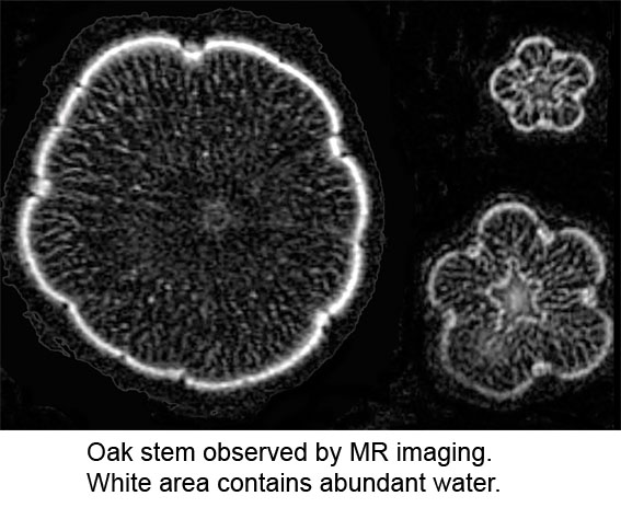
K., Kanbara, Y., Inoue, T. and Ogawa, A.: Magnetic resonance
micro-imaging of xylem sap distribution and necrotic lesions in tree
stems. IAWA Journal 27(1):3-17. 2006.
(PDF file download)
- Kuroda,
K., Ichihara,
Y., Kanbara,
Y.,
Inoue, T. and Ogawa, A.:
Visualization
of a host reaction in oak stems infected with a wilt pathogen, Raffaelea quercivora,
by magnetic resonance imaging. 6th Pacific
Regional Wood Anatomy Conference 2005 Kyoto Japan, Abstracts
p.64-65,
2005
- Kuroda,
K.: Xylem dysfunction in Yezo spruce (Picea jezoensis)
after
inoculation with the blue-stain fungus Ceratocystis polonica.
Forest
Pathology 35(5): 346-358. 2005. (PDF
file download)
- Kuroda, K.:
Inhibiting factors of
symptom development in several Japanese red pine (Pinus densiflora)
families selected
as resistant to pine wilt. Journal of Forest Research 9:
217-224,
2004
(PDF
file download)
- Kuroda, K.,
Ichihara,
Y., Kanbara, Y., Inoue, T. and Ogawa, A.: Magnetic
Resonance imaging
of xylem
dysfunction in Quercus
crispula
infected with a wilt pathogen, Raffaelea
quercivora.
Abstract of IUFRO Working Party 7.02.02 FOLIAGE, SHOOT
& STEM
DISEASES, June 13-19, 2004, Oregon, USA, P.16, 2004.
- Kuroda,
K.:
Degradation of Conifer Plantations in the Kansai
District. Bulletin of the Forestry and
Forest Products Research Institute, 2:247-254, 2003.
(PDF
file
download)
- Kuroda,
K., Kanbara,
Y., Inoue, T. and Ogawa, A.:
Analysis
of NMR-CT
images
to detect
the xylem dysfunction and lesions in tree trunks. Abstract of
5th
Pacific Regional Wood Anatomy Conference (Indonesia, Yogyakarta), IAWA
Journal, 23:469-470, 2002
- Kuroda, K.:
The
mechanism of tracheid cavitation in trees infected with wilt
diseases. Proceedings of the IUFRO Working Party 7.02.02 shoot and
Foliage Diseases, P.17-23, Hyytiala, Finland, 2001
- Kuroda, K.: Responses of Quercus
sapwood to infection with the pathogenic fungus of a new wilt
disease vectored by the barkbeetle Platypus quercivorus.
J.
Wood Science 47: 425-429 , 2001 (PDF
file download,
Abstract)
- Kuroda, K.: Anatomical assessment of age of
infection with resinous stem canker
in hinoki cypress
(Chamaecyparis obtusa) and the factors promoting resinosis. J. Jpn
Wood Res Soc, 46(6), 503-509, 2000
- Kuroda, K.
Kuroda, H. Lewis, A. M.:
Detection of embolism and acoustic emissions in tracheids
under a
microscope: Incidence of diseased trees infected with pine wilt. New
Horizons in wood Anatomy, ed. by YS Kim, Chonnam Nat'l Univ. Press,
Kwangju, Korea, 372-377, 2000 (The 4th Pacific Regional Wood Anatomy
Conference in Korea, 1998
- Kuroda, K.: Seasonal
variation in
traumatic resin canal formation in Chamaecyparis
obtusa
phloem. IAWA J. 19: 181-189, 1998
- Kuroda, K.
and Kiyono, Y.: Seasonal Rhythms
of Xylem Growth Measured by the Wounding Method and with a
Band-Dendrometer: An instance of Chamaecyparis Obtusa
. IAWA
Journal 18:291-299, 1997.
- Kuroda, K. and
Yamada, T.: Discoloration of
sapwood and blockage of xylem sap ascent in the trunks of
wilting Quercus
spp. following attack by Platypus quercivorus. J.
Jpn. For.
Soc. 78(1):84-88, 1996.
- Kuroda,
K.: Acoustic emission technique for the detection of abnormal
cavitation in pine trees infected with pine wilt disease. International
symposium on pine wilt disease caused by pine wood nematode (China).
Proceedings 53-58, 1996
(PDF
file
download)
- Kuroda, K.,
Yamada, T. and Ito, S.: Bursaphelenchus xylophilus
induced pine wilt: Factors associated with resistance. Eur. J. For.
Path. 21, 430-438, 1991 (PDF file
download)
- Kuroda, K.: Mechanism
of cavitation development in the pine
wilt disease. Eur. J. For. Path. 21, 82-89, 1991 (PDF
file download)
- Kuroda,
K.: Terpenoids causing tracheid-cavitation in Pinus
thunbergii infected by the pine wood nematode (Bursaphelenchus
xylophilus ). Ann. Phytopath. Soc. Jpn. 55, 170-178,
1989 (PDF
file download)
- Kuroda, K., Yamada, T. and Mineo, K. Tamura, H.: Effects of
cavitation on the development of pine wilt disease caused by
Bursaphelenchus xylophilus. Ann. Phytopath. Soc. Jpn. 54,
606-615,
1988
(PDF file download)

Abstracts
of publications: '96-2001
Keiko Kuroda:
Responses of Quercus sapwood to
infection with the
pathogenic fungus of a new wilt disease vectored by the barkbeetle Platypus
quercivorus. J. Wood Science 47: , 2001.
Quercus serrata and Q.
crispula wilt
during the summer in wide areas along the Sea of Japan. Mass attacks
of trees by an ambrosia beetle (Platypus quercivorus
) are
characteristic before the appearance of wilting symptoms. This study
investigated the pathogenic effects of a fungus detected specifically
in the wilting trees. This fungus, tentatively named Ooak fungus, has
a distribution that correlates the discolored xylem area called wound
heartwood, in which vessels were dysfunctional. Tylosis formation
around
the hyphae indicates the vessel dysfunction. In the areas under
discoloration, the fungal hyphae were invading living ray parenchyma
cells from vessel lumen. As a protective reaction, the ray cells
exuded yellow substances into the vessels. However, these substances
seemed ineffective against fungal activity, probably because the
fungus disperses along the beetle's gallery before enough substance
could accumulate. It should allow the wide discoloration in sapwood.
Cambium was not necrotic around the fungus. Cytological process in the
host was as follows: (1) Synthesis of secondary metabolites by the
stimuli of oak fungus, (2) exudation of yellow substances into vessels,
and (3) dysfunction of vessels and wound heartwood formation. In the
wilting incidence of trees, pathogenicity of the fungus should be
assessed by the ability to stop sap-flow.
Key words: Ambrosia beetle, Xylem discoloration, Wound
heartwood,
Vessel dysfunction, Secondary metabolites
Download of PDF file
Return to Recent
papers
Kuroda, K.:
Anatomical assessment of age of infection with resinous stem canker
in hinoki cypress (Chamaecyparis obtusa) and the factors promoting
resinosis. J. Jpn Wood Res Soc, 46(6), 503-509, 2000
Highly frequent traumatic resin canal
formation in
the phloem is characteristic of resinous stem canker disease. A
fungus, Cistella japonica, was reported as a candidate for causal
agent, and some environmental factors may affect extensive and
long-term resinosis in tree trunks. At several plantations in Maizuru
and Kanazawa in Japan, resin canal formation and resin production
appear
to be active under conditions that promote tree growth. In the
plantations where this disease was frequently observed, even trees
without resinosis contained traumatic resin canals. Such trees are not
healthy, but 'diseased trees without visible symptoms'. Extensive
resinosis is often discovered at mature plantations about 20 years old.
However, the onset of this disease was estimated to occur at the very
young age of about 5 to 8 years, based on the fact that wound resin
canals are not formed in the phloem more than three years old.
Disease-promoting factors should therefore be surveyed tracing back to
the earlier period. The distribution of resin canals throughout a trunk
circumference suggests that the stimuli that induce epithelial cell
differentiation affect the entire trunk. Some physiological aspects of
trees may relate to the sensitivity to resin canal formation. Partial
necrosis of cambium was observed in the specimens with resinosis,
except
for the youngest of age 11, and was associated with resin pockets.
Well-grown trees have a tendency to promote resinosis and cambium
necrosis.
Return to
Recent papers
Kuroda, K.
Kuroda, H. Lewis, A. M.:
Detection of embolism and acoustic emissions in tracheids under a
microscope: Incidence of diseased trees infected with pine wilt. New
Horizons in wood Anatomy, ed. by YS Kim, Chonnam Nat'l Univ. Press,
Kwangju, Korea, 372-377, 2000 (The 4th Pacivic Resional Wood Anatomy
Conference in Korea, 1998, IAWA Journal 19(4) P.463-464 1998).
Xylem-sap in the water conduits is kept
under
tension when transpiration is active. The conduit's water columns can
break under high tension and form bubbles (emboli). In healthy
plants, water columns recover by rehydration when the tension is
reduced. In trees infected with wilting diseases, however, sap ascent
finally stops without recovering in dehydrated xylem areas. We observed
embolism in light-microscope sections of diseased trees and confirmed
the relationship between bubble development and acoustic emissions
(AEs)
that are detected at embolism. We discuss the mechanism of water
blockage in pine wilt disease.
Three-year-old
Japanese red-pines (Pinus densiflora), inoculated with pine wood
nematodes (Bursaphelenchus xylophilus) and healthy trees, were used.
Embolism was observed on radial sections (1x6mm) of 60 µm
thick that
contain a layer of intact tracheids, and was recorded on videotape
(Lewis' method). At the same time, AEs were monitored with an
AE-transducer attached to the sections. As the second experiment, the
time necessary for the rehydration of healthy and infected pines
following the addition of water was compared.
First,
dehydration without AEs occurred from cut-ends of tracheid injured
during sectioning. Then, bubbles emerged near the centers of intact
tracheids, abruptly swelled, and filled whole tracheids. Such bubble
expansion is thought to occur by the evaporation of water into a very
tiny bubble. During high-rate bubble formation, AEs were produced. We
successfully recorded AEs as audible-sound through the audio terminal
of the VTR. The AEs coincided with almost all of the rapid bubble
development. This result supports the idea that AEs detected in the
trunks of living trees are produced by embolism in tracheids. Two weeks
after inoculation of the pathogen, water blockage by embolism had just
occurred in a part of the xylem. In such trees, the time necessary for
rehydration is longer than in healthy trees. It suggests that certain
substances that inhibit bubble dissolution may exist in xylem.
In detail, see
movie
Return to Recent
papers
Keiko
Kuroda:
Seasonal
variation in traumatic resin canal formation in Chamaecyparis
obtusa
phloem. IAWA J. 19: 181-189, 1998.
Trunks of Chamaecyparis obtusa were injured to
examine the
seasonal differences in traumatic resin canal formation in secondary
phloem. Even after the wounding during winter, differentiation of axial
parenchyma into epithelium was initiated, and vertical resin canals
formed. After winter wounding, resin canal development was slower, the
tangential extent of resin canals was narrower, and it took one to two
months until resin secretion began. After spring wounding, the sites
of resin canal formation were the one- and 2-year-old annual ring of
phloem. In August, the location shifted into the current and
one-year-old annual ring. Resin canals never formed in areas that were
3
or more years old. In C. obtusa trunks that are affected by the
resinous
stem canker, numerous tangential lines of resin canals are found
throughout the phloem, not just recent and 1--2 year old phloem. The
present research indicates that these many lines of resin canals were
not formed at one time, and that the stimuli that induce traumatic
resin canals must occur repeatedly over many years. The data on
artificial wounding effects are useful for understanding resinous stem
canker.
Key words: Traumatic resin canal, secondary phloem, Chamaecyparis
obtusa, resinous stem canker, injury
Return to
Recent papers
Keiko
Kuroda and Yoshiuki
Kiyono:
Seasonal Rhythms of Xylem Growth Measured by the Wounding Method and
with a Band-Dendrometer: An instance of Chamaecyparis Obtusa.
IAWA Journal 18:291-299, 1997.
The pinning method for the
measurement of xylem
growth was modified for easier application. Trunks of Chamaecyparis
obtusa were monthly incised with a knife instead of a thin
needle.
Two years later, xylem blocks including wounded areas were all
harvested, and xylem growth curves for two years were reconstructed
from the sites of wound tissue. Circumferential increases were measured
with the band-dendrometer on the same trees for comparison.
Measurement by wounding method indicated a tendency for cambial cell
production to accelerate twice a year, around May and August.
Circumferential increase measured with the band dendrometer differed
from radial growth. It was very small around August and continued after
the cessation of cell-production. The climatic data near the plantation
suggested circumferential size of trunks is probably affected by the
physical shrinkage of trunks because of water shortage during the
drought season and trunk swelling following precipitation.
Circumferential increments did not reflect the seasonal rhythms of
xylem growth. Therefore, for the detailed information on the radial
growth within a season, the wounding method is recommended. Key
words: Xylem growth, pinning method, wounding method, band-
dendrometer, drought shrinkage, Chamaecyparis obtusa
.
Return to
Recent papers
K.
Kuroda and T. Yamada:
Discoloration of sapwood and blockage of xylem sap ascent in the
trunks of wilting Quercus spp. following attack by
Platypus
quercivorus. J. Jpn. For. Soc. 78:84-88, 1996.
Many deciduous oak trees, Quercus
serrata
and Q. crispula are wilting during summer in the
wide areas of
Honshu island of Japan along the Japan sea. Such forests that had
been used for charcoal production are not managed appropriately now.
Prior to wilting, mass attacks by an ambrosia beetle, Platypus
quercivorus , into trunks were observed. A specific fungus,
which
is carried into xylem via beetles' mycangia, had been detected from
wilting trees. We discussed on the determinant factor of this oak
wilting, from the observation of tree tissues taken from trees attacked
by the beetle. Following the beetles' invasion, xylem discoloration had
occurred in sapwood surrounding long galleries whether the tree is
wilting or not. Fungal hyphae were found in vessels near the
galleries. The xylem sap ascent was blocked at such discolored xylem
like heartwood. Discolored area became maximum where beetles' gallery
elongated in high density usually from the base to the breast height
of trunks. Before the start of wilting symptom, sap ascent had been
completely blocked in trunks at the height of maximum discoloration for
most vessels became dysfunctional in the current annual ring, and
occasionally cambium was necrotic.
Return to top


